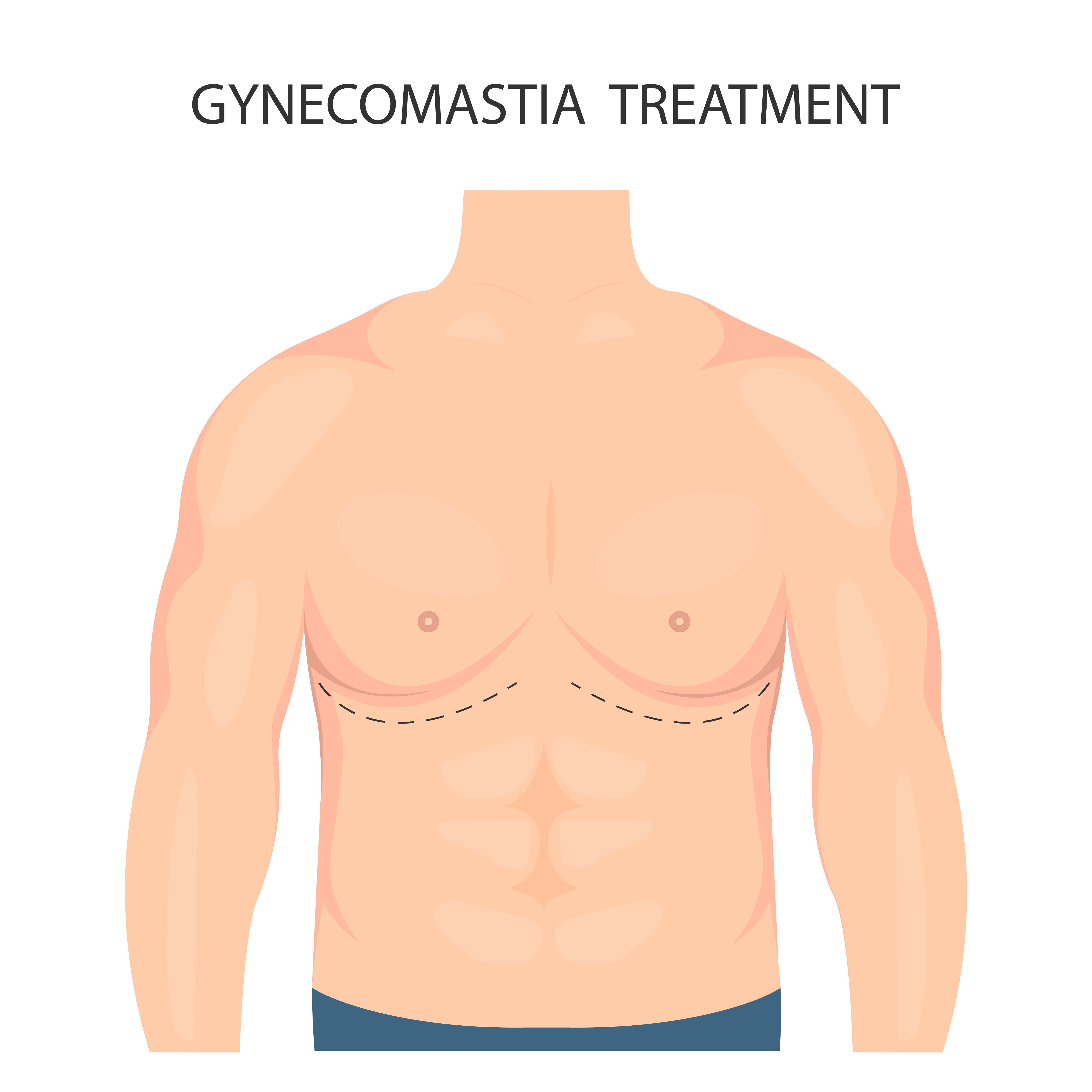
A larger male chest can result from soft fat, a firm gland beneath the nipple, loose skin, or a combination of these features. By the end, you will know what an examination, medical work-up, severity grade, and tissue-specific operation each contribute to a safe treatment plan.
Key takeaways
- Firm tissue behind the nipple points to gland enlargement, not chest fat alone.
- Treat puberty, medicines or underlying conditions before planning surgery.
- Simon grade and skin laxity help determine whether skin removal is needed.
- Liposuction removes fat; excision removes firm gland tissue; many cases need both.
How the examination separates fat, gland and warning signs
The key distinction is the tissue you can feel, not the overall chest size. A surgeon examines the chest while standing and lying down, comparing both sides for softness, firmness, mobility and the location of the enlargement.
| Pattern | Examination finding | Treatment implication |
|---|---|---|
| pseudogynecomastia | Soft, diffuse fat without a defined central lump | Weight loss may reduce it; liposuction can contour persistent fat |
| true gynecomastia | A firm disc beneath the nipple-areola complex, often rubbery and mobile | Dense glandular tissue may need direct subareolar excision |
| mixed gynecomastia | Soft surrounding fat plus a firm central gland | Combined liposuction and gland excision may be needed |
The examination also records whether the tissue is centred under the areola or spread across the chest, along with nipple position, skin elasticity, asymmetry, loose skin and the shape of the underlying muscle.
These details prevent a plan based only on chest volume: weight loss can reduce fat but does not reliably remove established glandular tissue.
A hard, fixed or eccentric lump, nipple retraction, discharge, skin tethering or enlarged axillary nodes is not a routine cosmetic pattern. Those findings warrant medical investigation, and excised tissue is commonly sent for histopathology when enlargement is firm, one-sided or otherwise atypical.
The goal is balanced contour, not removal of every structure beneath the nipple; taking everything can create a visible crater, while leaving excess gland can preserve puffiness.
When medicines, health conditions or puberty change the plan
Chest enlargement should receive medical assessment before cosmetic treatment when it is recent, painful, rapidly increasing, asymmetric, or linked to a medicine, substance, or health change. Operating first can miss a reversible cause and leave the enlargement untreated.
1. Review every medicine and substance. Tell the clinician about spironolactone, finasteride, anabolic steroids, cannabis, alcohol, antidepressants, antipsychotics, heart medicines and antiandrogens. Do not stop a prescribed drug without the prescriber’s advice; the safer plan may be substitution or treatment of the underlying condition.
2. Investigate possible hormone or organ disorders when enlargement is unexplained, severe or persistent. Assessment may include examination of the testes and thyroid, plus blood tests for liver function, kidney function, testosterone, estradiol, luteinising hormone and human chorionic gonadotropin. The exact tests depend on the findings.
3. Seek prompt assessment for a hard or fixed lump, a mass away from the nipple, nipple retraction, discharge, skin tethering or enlarged armpit nodes. These findings do not fit ordinary pubertal or medication-related gynecomastia and require investigation before cosmetic planning.
4. Treat adolescent enlargement differently. Puberty-related gynecomastia often regresses, so observation is appropriate when examination is reassuring. Persistent, severe or painful enlargement after physical development stabilises can justify a surgical discussion, especially when distress remains substantial.
Recent painful tissue is more likely to respond to specialist-directed treatment such as tamoxifen than long-standing, fibrotic tissue. Medication is not a universal substitute for surgery, and its evidence and regulatory status vary by setting.
How Simon grade and loose skin affect the operation
Simon classification helps estimate how much enlargement and skin excess must be addressed, but it does not prescribe the operation. Simon grade I means small enlargement without excess skin; Simon grade IIa means moderate enlargement with a tight skin envelope; Simon grade IIb means moderate enlargement with excess skin.
- Simon grade I: removing glandular tissue, fat, or both can be enough when the nipple sits normally and the skin recoils well.
- Simon grade IIa: tissue removal often remains sufficient, although a broad chest or low nipple can require additional contour planning.
- Simon grade IIb: removing tissue alone can leave a loose, folded envelope, so skin reduction may be discussed.
- Simon grade III: marked enlargement with excess skin commonly requires a larger skin-removal and nipple-repositioning plan.
The examination matters more than the label. The surgeon checks breast width, skin elasticity, nipple descent, asymmetry and whether the nipple sits below the chest’s natural contour. A low or downward-pointing nipple signals that volume reduction alone could leave an empty, drooping envelope.
Keeping the skin avoids longer scars and reduces risks such as wound separation, numbness and impaired nipple blood supply. Removing it improves the hanging outline but creates visible scars and may require nipple grafting or repositioning in severe cases.
The goal is a flat, even contour, not removal of every structure beneath the areola; over-excision can create a crater deformity.
Why the tissue pattern determines liposuction, excision or both
A soft, diffuse chest enlargement is usually treated with liposuction, while a firm disk beneath the areola points toward subareolar gland excision. Mixed enlargement often needs both, because liposuction removes fat but cannot reliably flatten dense glandular tissue.
| Option | Tissue pattern | Typical approach | Main trade-off |
|---|---|---|---|
| Liposuction | Predominantly fatty enlargement | Remove fat through small access points | Smaller scars, but residual gland can leave a visible mound |
| Subareolar gland excision | Firm, concentrated gland beneath the nipple | Remove the gland directly through an areolar incision | Better control of the disk, but adds scar, swelling and contour risks |
| Combined treatment | Fat plus gland | Liposuction followed by gland excision | More complete contour correction, with more operative time and recovery |
The decision should follow tissue distribution, not chest size alone. Losing weight can reduce fat in pseudogynecomastia, but it will not reliably remove established glandular tissue; treating a lean patient with liposuction alone can therefore leave the nipple area prominent.
1. Ask how much gland will remain after fat removal. A small residual layer can prevent a hollow appearance, but too much leaves fullness; removing too aggressively can create a crater or an uneven areola.
2. Discuss skin quality before choosing a larger operation. Mild laxity may contract after tissue removal, whereas hanging or redundant skin may require skin removal and nipple repositioning.
3. Balance correction against scars and sensation. Skin removal can improve a loose envelope, but creates longer, more visible scars and raises the risk of altered nipple sensation, wound problems or reduced nipple blood supply.
4. Match the plan to your priorities: the flattest contour, the shortest scars, the lowest chance of residual tissue or tolerance for staged correction.
What an individualized treatment discussion should cover
Before consenting, confirm the diagnosis, the reason for each test, the tissue plan and the result you are realistically being promised. Imaging is not automatic: an indeterminate examination may justify breast ultrasound, while a hard, fixed, eccentric mass, nipple discharge or skin tethering may require diagnostic mammography or biopsy assessment.
Ask these questions in the consultation:
- Which findings support fat, gland or both, and what would change the operation? Ask whether your history warrants testosterone, estradiol, LH and FSH, plus liver and kidney function tests. These tests investigate hormonal or systemic causes; they are not an identical checklist for every patient.
- Will removed tissue be sent for histopathology, and when will you receive the report? A firm, unusual or one-sided lesion deserves a clear pathology plan rather than being treated as routine tissue.
- Where will the incisions sit, how long will scars remain noticeable, and could nipple sensation or position change? Confirm whether loose skin requires excision, because removing volume alone can leave folds or a drooping nipple.
- What contour, asymmetry, hematoma, seroma, infection, altered sensation or revision risks apply to you? Ask how much gland will remain beneath the areola; leaving a thin layer can prevent a crater but can also leave fullness.
- What could cause residual or recurrent enlargement? Persistent hormone or drug causes, substantial weight change or untreated medical disease can undo a technically good result.
Whether you consult Dr Nakul Somani or another surgeon, request expectations in plain terms: improvement, not guaranteed symmetry or permanent zero volume.
Related service
Cosmetic Surgeries Gynecomastia Get Rid of Excess Fat in Your Chest by Undergoing Male Breast Reduction/Gynecomastia at Engrace Clinics Due to an excess storage of fat around the... |
Frequently asked questions
How does an examination distinguish fat from glandular gynecomastia?
The surgeon examines you standing and lying down, comparing both sides for softness, firmness, mobility and the location of enlargement. Soft, diffuse tissue suggests fat, while firm tissue behind the nipple suggests gland enlargement.
When can medicines, health conditions or puberty change the treatment plan?
Recent puberty-related enlargement, medicines and health conditions can cause or worsen gynecomastia. The cause should be assessed and treated before surgery when appropriate.
How do Simon grade and loose skin affect gynecomastia surgery?
Simon grade describes the amount of enlargement and skin excess. More enlargement or loose skin can require tissue excision or skin removal in addition to liposuction.
When is liposuction, excision or combined surgery used?
Liposuction targets soft fatty tissue. Excision removes firm gland tissue, usually through an incision near the areola. Combined surgery addresses both fat and gland enlargement.
What should an individualized gynecomastia treatment discussion cover?
Discuss the tissue pattern, likely cause, Simon grade, skin quality, procedure options, scars, asymmetry, risks, recovery and the result you consider realistic.